Animal Cell Under The Microscope - Eukaryotic Animal Cell Under Microscope - Micropedia : 15 видео 74 483 просмотра обновлен 16 апр.
byLeon Avarbuch-0
Animal Cell Under The Microscope - Eukaryotic Animal Cell Under Microscope - Micropedia : 15 видео 74 483 просмотра обновлен 16 апр.. (reproduced by permission of photo. The lesson will also take you through some exam questions on finding magnification using a scale and an image of a cell. The differences between plant and animal cells. Animal cells are of various sizes and have irregular shapes. As per the given information in the question, cells of mushrooms, plants, and animals all have visible nuclei under a microscope.
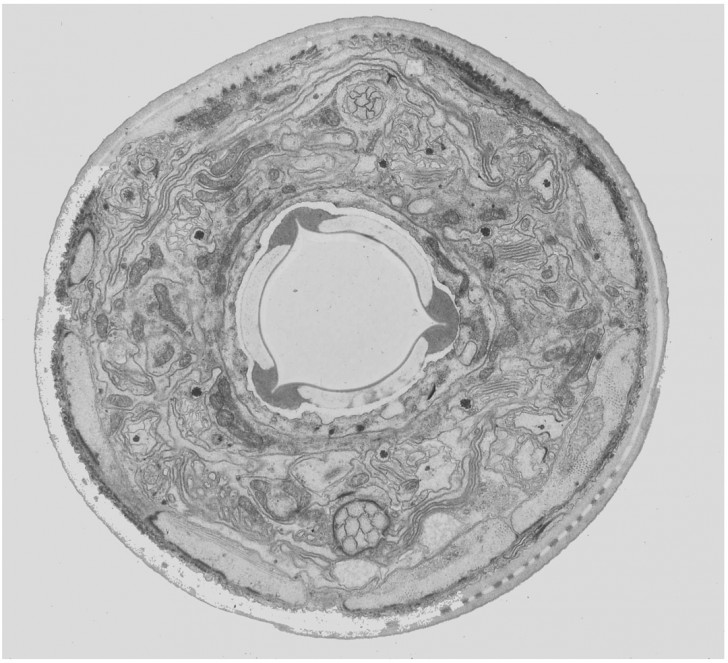
A generalised animal cell as observed under an electron microscope. Some features common to animal cells. The differences between plant and animal cells. In this video, you will explore 3 different microscopic views of human. It also has a very high resolving power.
animal cells under a microscope | Under the microscope ... from s-media-cache-ak0.pinimg.com Place the glass slide onto the stage. A cell is a very tiny structure which exists in living bodies. Cells consist of cytoplasm enclosed within a membrane, which contains many biomolecules such as proteins and nucleic acids.2 most plant and animal cells are only visible under a light microscope, with dimensions between 1. Plant cells have cell walls, one large vacuole per cell, and chloroplasts, while animal cells will have a cell membrane only. Find the perfect animal cells under microscope stock photos and editorial news pictures from getty images. Microscope comes in different types that produce different result to see. Observe the onion skin under low power of the microscope and then under high power. You will know if a cell if a plant or animal by looking under a microscope.
Under a light microscope, the cell membrane, nucleus and cytoplasm of a cheek cell (animal cell) can be observed.
Cells consist of cytoplasm enclosed within a membrane, which contains many biomolecules such as proteins and nucleic acids.2 most plant and animal cells are only visible under a light microscope, with dimensions between 1. When we look at cells under the microscope, our usual measurements fail to work. Most of the cells size range between 1 and 100 micrometers and are visible only with the microscope. We can view animal cells using a microscope and dye under low and high power magnification. Elle means small or minor, so organelles are basically minor, microscopic organs. Animal cells are of various sizes and have irregular shapes. Cell biology (also called cellular biology or cytology) is the study of cells. Under a light microscope, the cell membrane, nucleus and cytoplasm of a cheek cell (animal cell) can be observed. Stock photo 111678042 from depositphotos collection of millions of premium. It also has a very high resolving power. Digital artwork creative graphic design. 15 видео 74 483 просмотра обновлен 16 апр. The animal cell is more.
Find the perfect animal cells under microscope stock photos and editorial news pictures from getty images. The images were acquired using an andor revolution spinning disk system with an olympus microscope. Each of these epithelial cells was examined under the microscope as students. Select the lowest power objective lens. To look at a cell close up we need a microscope.
animal cell microscope : Biological Science Picture ... from pulpbits.net 7 ultrastructure of an animal cell as seen through an electron microscope. Professor griffiths is a wellcome trust principal research fellow. Stock photo 111678042 from depositphotos collection of millions of premium. Examining plant cells under the microscope. Eucalyptus, picture of eucalyptus, micro picture of cell structure of a plant, section cut microscopic nature, closeup plant life , microscope, mikroskopisch. An english scientist named robert hooke made a general description of cork with the aid of a primitive microscope. Image:plant cell seen under electron microscope. To look at a cell close up we need a microscope.
Image:plant cell seen under electron microscope.
To look at a cell close up we need a microscope. Elle means small or minor, so organelles are basically minor, microscopic organs. Most of the cells size range between 1 and 100 micrometers and are visible only with the microscope. Resolving power is the ability to distinguish between separate things which are close to each other. Some features common to animal cells. Under the microscope, an animal cell shows many different parts called organelles, that work together to keep the cell functional. Cells consist of cytoplasm enclosed within a membrane, which contains many biomolecules such as proteins and nucleic acids.2 most plant and animal cells are only visible under a light microscope, with dimensions between 1. Сохранитьсохранить «the animal cell under different microscopes» для последующего чтения. 15 видео 74 483 просмотра обновлен 16 апр. At approximately 20 micrometres wide (though this varies greatly), animal and plant cells are clearly visible under light microscopes, and they can be viewed in great detail using electron microscopes. It also has a very high resolving power. Digital artwork creative graphic design. The differences between plant and animal cells.
It also has a very high resolving power. Image:animal cell seen under electron microscope. 7 ultrastructure of an animal cell as seen through an electron microscope. Most of the cells size range between 1 and 100 micrometers and are visible only with the microscope. Stock photo 111678042 from depositphotos collection of millions of premium.
What does an animal cell look like under an electron ... from qph.fs.quoracdn.net In science, the metric system is used to measure objects and, as you will see, is vastly superior to our antiquated english system of there are three structures that distinguish plant cells from animal cells. In this video, you will explore 3 different microscopic views of human. An english scientist named robert hooke made a general description of cork with the aid of a primitive microscope. Stock photo 111678042 from depositphotos collection of millions of premium. It also has a very high resolving power. Most of the cells size range between 1 and 100 micrometers and are visible only with the microscope. (reproduced by permission of photo. Each of these epithelial cells was examined under the microscope as students.
It also has a very high resolving power.
The lesson will also take you through some exam questions on finding magnification using a scale and an image of a cell. Elle means small or minor, so organelles are basically minor, microscopic organs. Animal cells are of various sizes and have irregular shapes. As per the given information in the question, cells of mushrooms, plants, and animals all have visible nuclei under a microscope. The images were acquired using an andor revolution spinning disk system with an olympus microscope. In scanning microscopes the sample is illuminated with a bright well focused spot scanning over the sample. Each of these epithelial cells was examined under the microscope as students. Examining plant cells under the microscope. (reproduced by permission of photo. Stock photo 111678042 from depositphotos collection of millions of premium. Under the microscope, an animal cell shows many different parts called organelles, that work together to keep the cell functional. Digital artwork creative graphic design. 15 видео 74 483 просмотра обновлен 16 апр.


Post a Comment